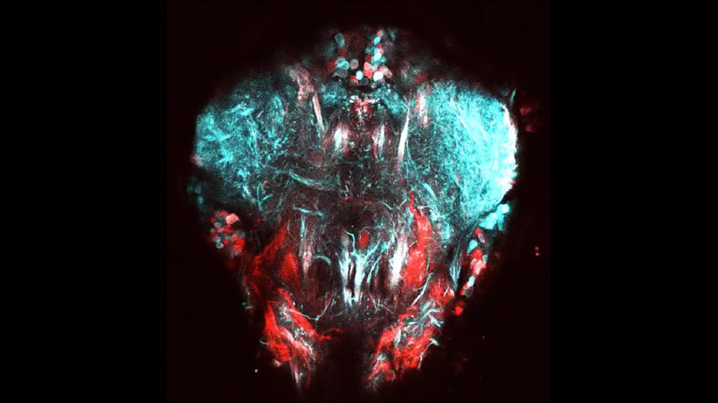
Scientists from the lab of Pavan Ramdya at EPFL’s Brain Mind Institute and Interfaculty Institute of Bioengineering have developed a new method for recording the activity of limb control neural circuits in the popular model organism, the fruit fly Drosophila melanogaster. The method uses an advanced imaging technique called “two-photon microscopy” to observe the firing of fluorescently labeled neurons that become brighter when they are active. wo-photon image of neural tissue controlling the front legs of t

Media properties
- Themes
- Research and Tech Transfer , SV, Robotics, Biology, Research and Tech Transfer
- Copyright
- EPFL - Pavan Ramdya
- Licence
- CC0 Licence
- Shooting date
- Oct. 22, 2018
- Album
- New technique reveals limb control in flies – and maybe robots
Share on