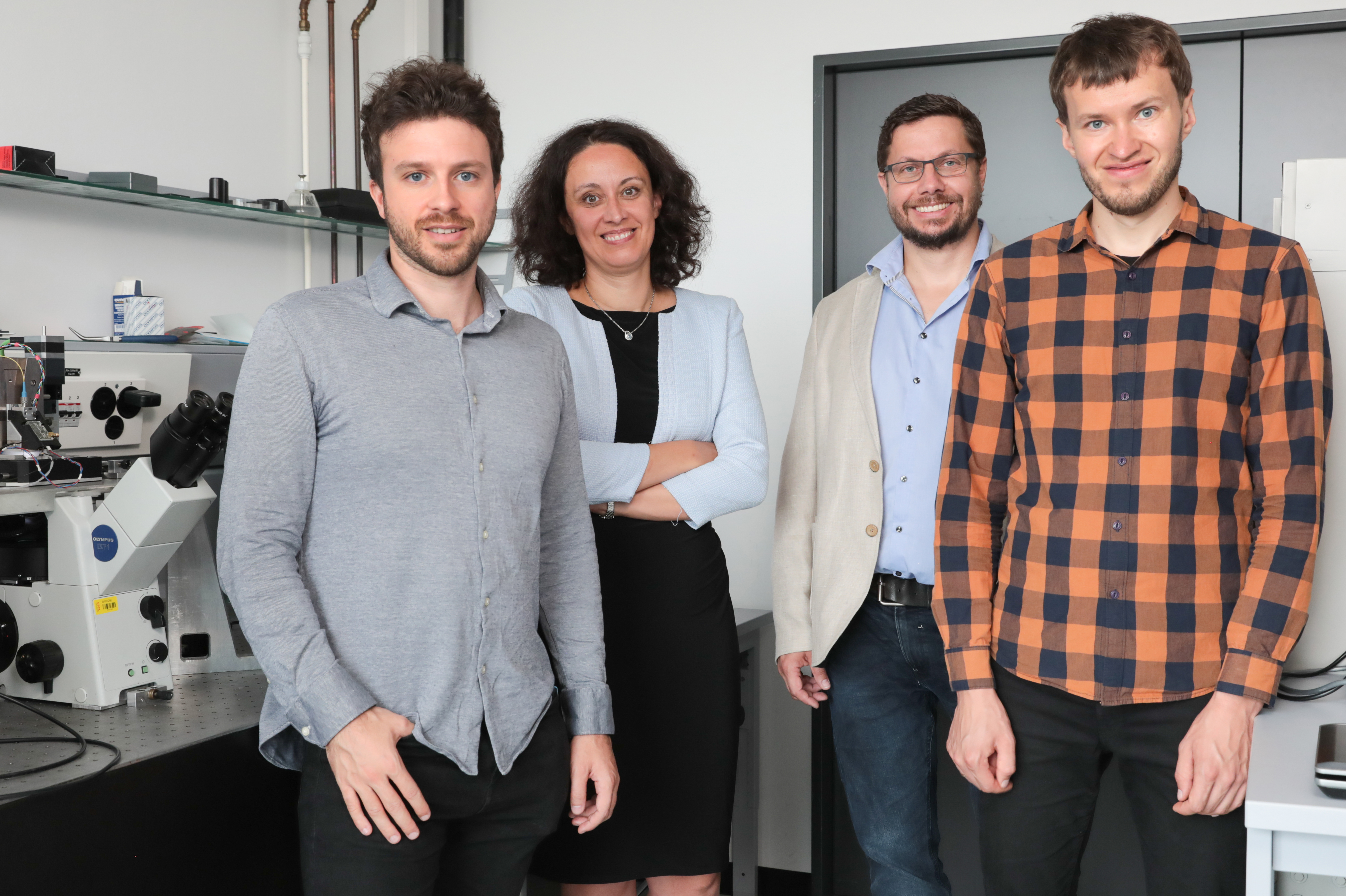
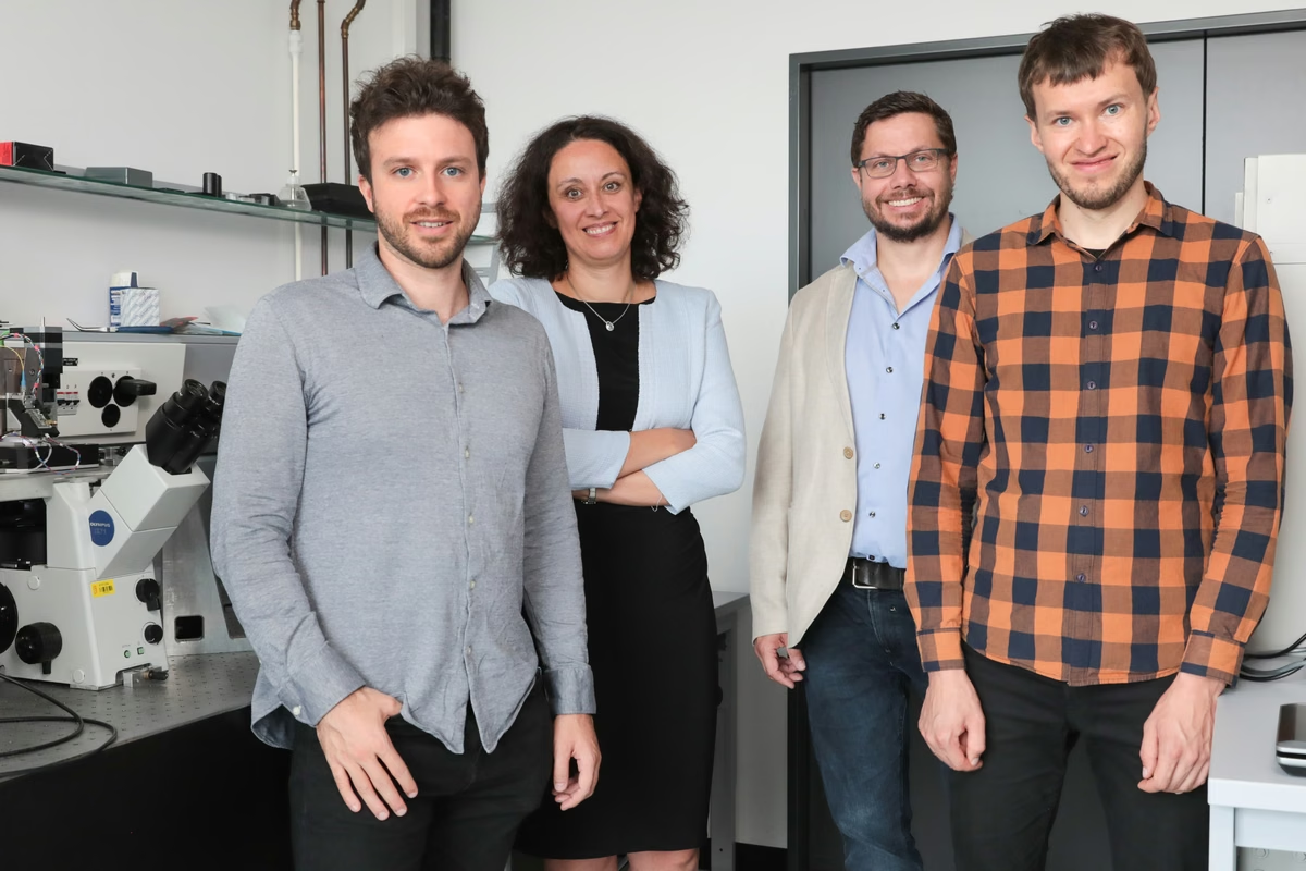
By combining two microscopy methods, EPFL researchers are able to see what is happening inside a cell and on its membrane simultaneously, giving unprecedented insight into the cellular processes that occur during infection, for example. From left to right: Samuel Leitao, doctoral assistant at LBNI, Aleksandra Radenovic, LBEN professor, Georg Fantner, LBNI professor, and Vytautas Navikas, doctoral assistant at LBEN.
Media properties
- Themes
- Research and Tech Transfer , STI, Microengineering, Biology, Research and Tech Transfer
- Copyright
- EPFL - Alain Herzog
- Licence
- CC0 Licence
- Shooting date
- Nov. 10, 2021
- Album
- New imaging method gives live glimpse into how cells work
Share on